– March 6, 2019
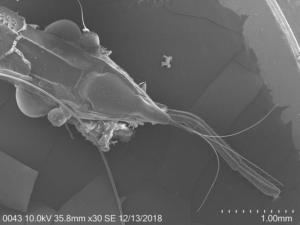
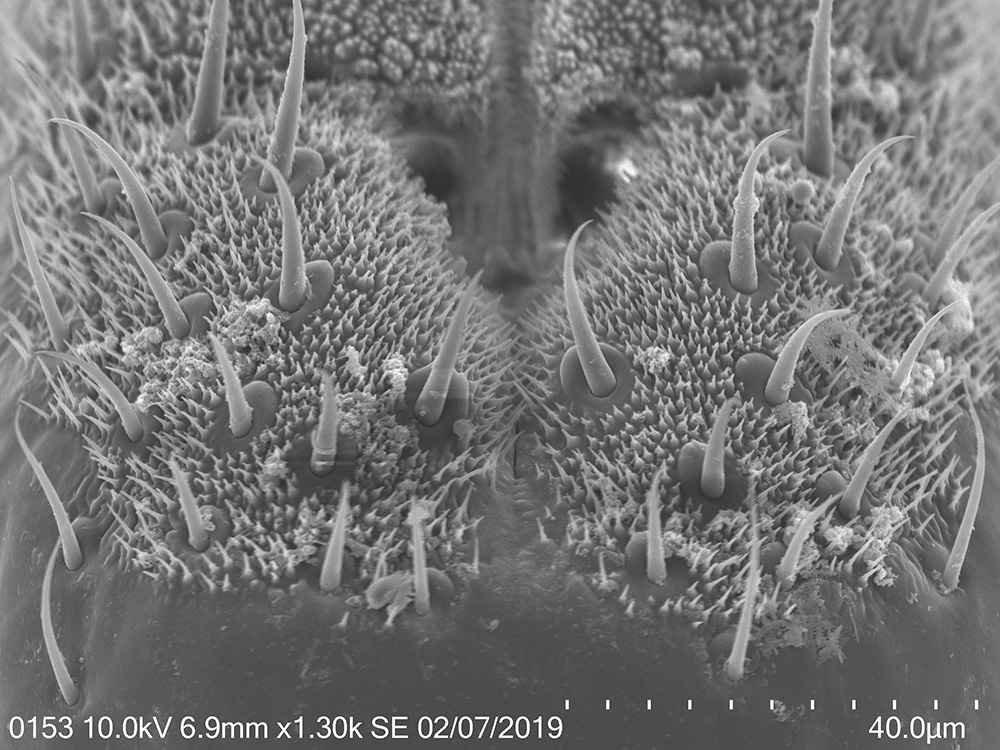
I’ve finished the 2nd round of scanning electron microscopy of the lanternfly mouthparts and tarsi which I conducted in The Laboratory for Biological Ultrastructure (my special thanks to Tim Maugel for his continuous help!!). We received beautiful SEM images, most of them are included now in my poster for the upcoming EB-ESA meeting. These images helped us to explore how different structures of the lanternfly mouthparts and tarsi change during the lanternfly development, and I’m very excited to start working on our new paper on the external morphology of the spotted lanternfly in relation to its host plants.
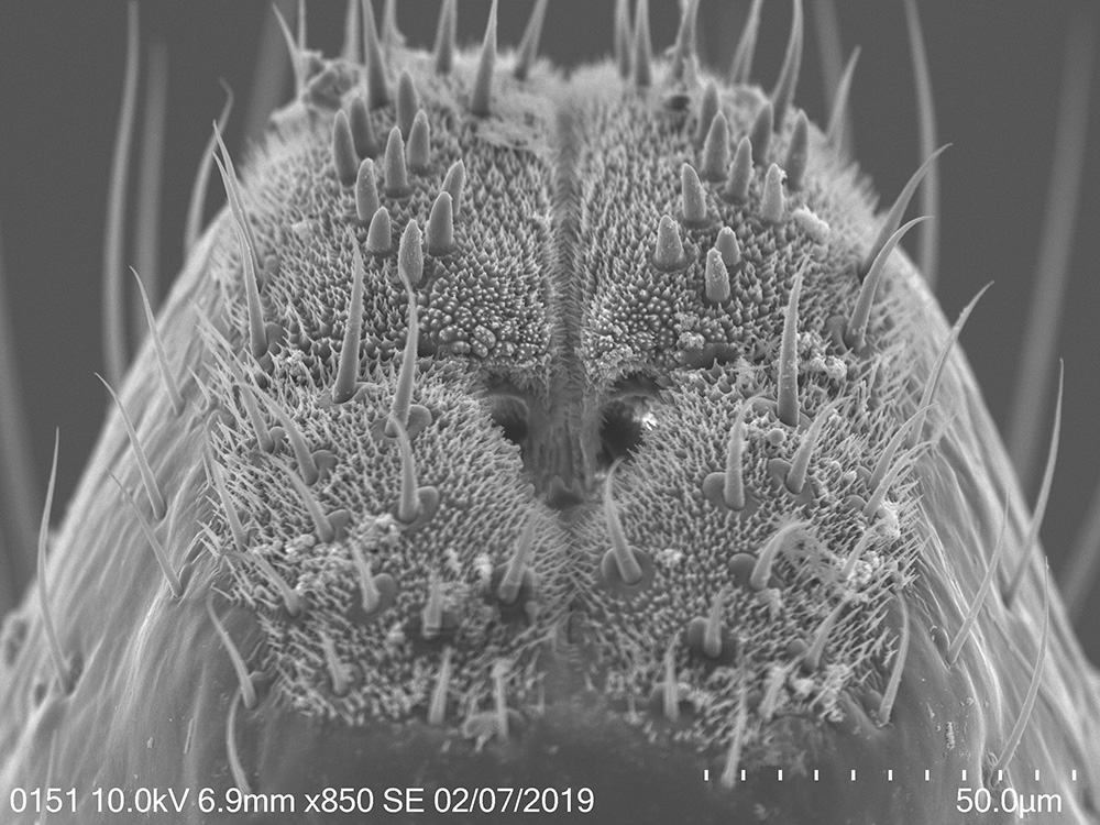
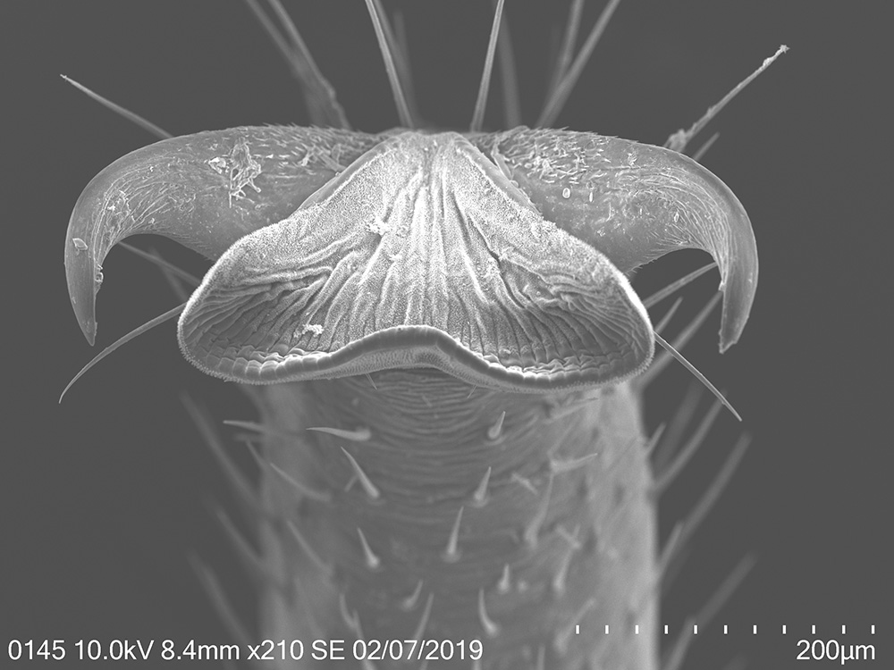
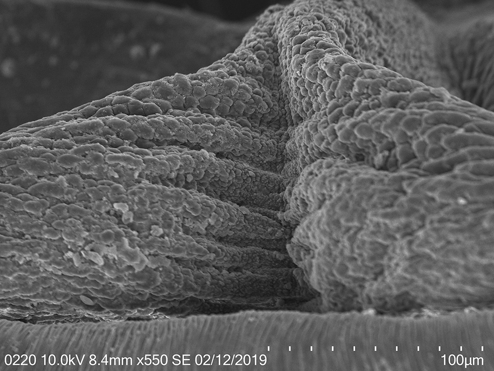
In this study we specifically focused on the structures which are responsible for primary contact of the spotted lanternfly with host tree surface (i.e. stylets, labium, tarsal claws, and arolia). We observed several interesting patterns:
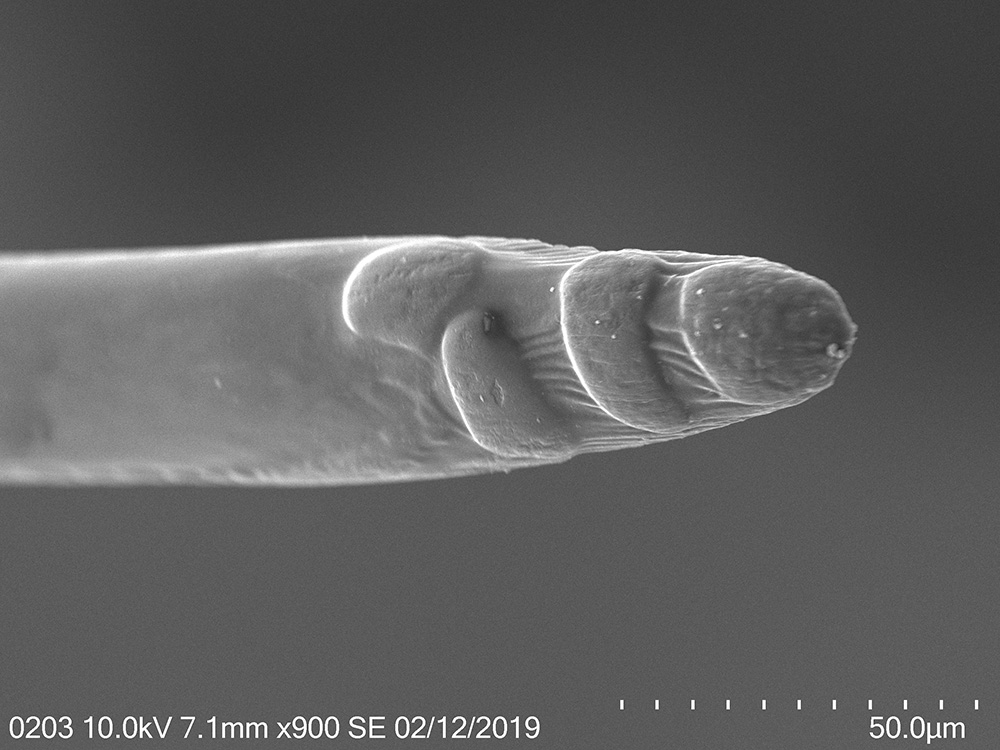
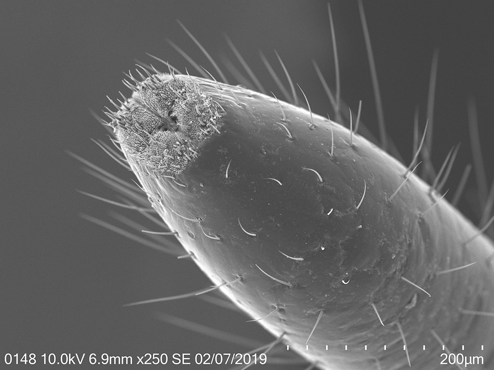
Mouthparts - oval prominences and longitudinal striations on the mandibular stylets are more defined in 4th instar and adults; labium is more segmented in adults; numerous sensilla, including the bristle-like sensilla, on the tip of the labium are present at each developmental stage.
Tarsal tips - tarsal claws are more spread out in adults; arolia are fully developed at each stage; arolia surface becomes more wrinkled in adults.
We think that observed patterns are adaptations to host plant usage and correspond with a type of host plants utilized at each developmental stage, and we would like to focus on morphometric comparisons of mouthparts and tarsi next. Can’t wait for these new results!