One year of work, four months in review, two months in revision, and here you go - a wonderful Christmas present from PLOS ONE! This paper summarizes the results of my morphological work on the spotted lanternfly, an aggressive invasive insect in the eastern US. In part, thanks to intensive training in scanning electron microscopy at the Laboratory for Biological Ultrastructure. In this study, I was able to investigate the external structures of the lanternfly mouthparts and tarsal parts, as well as a number of sensilla, which play an important role in the lanternfly primary contact with the surface of a host plant.
Avanesyan, A., Maugel T.K., and W. Lamp (2019) External morphology and developmental changes of tarsal tips and mouthparts of the invasive spotted lanternfly, Lycorma delicatula. PLOS ONE, doi.org/10.1371/journal.pone.0226995
A few selected figures:
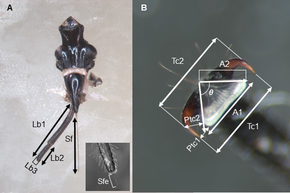
Morphometric characteristics measured for the labium, the stylet fascicle and the tarsal tip of Lycorma delicatula.(A) Labium and stylets. (B) Tarsal tip, the dorsal view. https://doi.org/10.1371/journal.pone.0226995.g001
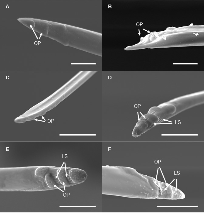
SEM of the mandibular stylets of Lycorma delicatula at each developmental stage. (A) First instar nymph. (B) Second instar nymph. (C) Third instar nymph. (D) Fourth instar nymph. (E) Adult female. (F) Adult male. https://doi.org/10.1371/journal.pone.0226995.g010
SEM of the tarsal tips of the forelegs of Lycorma delicatula at each developmental stage. (A) First instar nymph. (B) Second instar nymph. (C) Third instar nymph. (D) Fourth instar nymph. (E) Adult female. (F) Adult male. https://doi.org/10.1371/journal.pone.0226995.g012
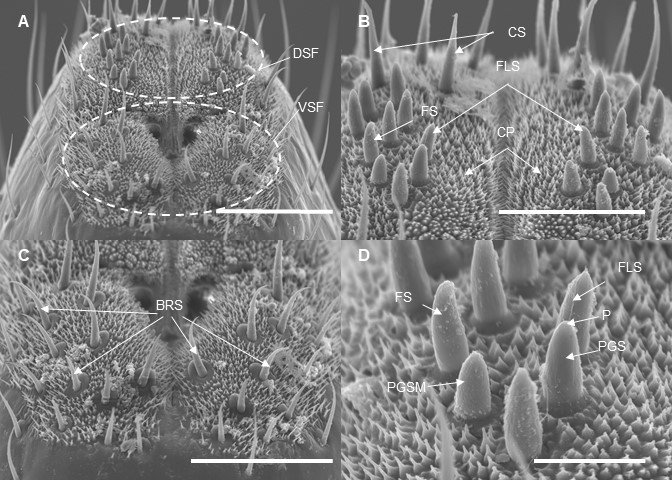
SEM of the labial tip of Lycorma delicatula (Third instar nymph). (A) Sensory fields. DSF, dorsal sensory field; VSF, ventral sensory field. (B) and (D) Dorsal sensory field. CS, clavate sensilla; FS, forticate sensilla, FLS, finger-like sensilla; CP, cuticular process; PGSM, multiporous peg sensilla; PGS, peg sensilla; P, pore. https://doi.org/10.1371/journal.pone.0226995.g008
It has been challenging and very interesting work. In this study, we focused on assessing changes in morphology of (a) the lanternfly mouthparts (stylets and labium), and (b) the lanternfly tarsal tips (arolia and tarsal claws) at each developmental stage. Our study revealed several interesting developmental patterns which potentially allow L. delicatula to better attach to a host plant and deeper penetrate to the host plant tissues at the late nymphal stages and adult stage. Here they are:
The labium in adults consists of five segments whereas the nymphs have four labial segments
The labium and stylet length, as well as the tarsal claw dispersal from the arolium, exponentially increase by 4th- instar nymph and adult stage
Mandibular stylets possess four indentations on the outer surface of the stylet apical part which become more evident in 4th-instar nymphs and adult
Longitudinal striations between indentations are present on mandibular stylets of 4th-instar nymphs and adults
Arolia surface becomes wrinkled in late instars and adults
Changes in the angle of arolium growth follow the quadratic growth curve; the angle becomes obtuse in adults which potentially causes increase of the arolia basal width
Additionally, we have found six morphological types of sensilla which are present at the labial tip at each developmental stage of L. delicatula; which potentially indicates the lanternfly ability to effectively explore the host plant suitability at each developmental stage.
This is the first study on variation in the lanternfly external morphology across various developmental stages, and our findings are critical for investigating and predicting the lanternfly host range, and the lanternfly dispersal to new host trees at each developmental stage.